Table des Matières
Publicité
Les langues disponibles
Les langues disponibles
Liens rapides
Publicité
Chapitres
Table des Matières
Dépannage

Manuels Connexes pour Optika Italy OPTIGEM Serie


Sommaire des Matières pour Optika Italy OPTIGEM Serie
- Page 1 OPTIGEM Series INSTRUCTION MANUAL Model OPTIGEM-10 OPTIGEM-20 Ver. 1.0 2021...
-
Page 2: Table Des Matières
Table of contents Warning Safety Information Package content OPTIGEM 10 OPTIGEM 20 Unpacking Intended use Symbols and conventions Instrument description OPTIGEM 10 OPTIGEM 20 Assembling Use of the microscope Adjusting interpupillary distance Focusing Adjusting the tension of the focus knob Diopter compensation 9.5 Magnification... -
Page 3: Warning
Warning This microscope is a scientific precision instrument designed to last for many years with a minimum of maintenance. It is built to high optical and mechanical standards and to withstand daily use. We remind you that this manual contains important information on safety and maintenance, and that it must therefore be made accessible to the instrument users. -
Page 4: Package Content
Package content OPTIGEM 10 ① ② ③ ⑤ ④ ⑥ ① Microscope stand ④ Jewel clip ② Binocular microscope body ⑤ Dust cover ③ Eyepieces ⑥ Power supply Page 4... -
Page 5: Optigem
OPTIGEM 20 ① ② ③ ⑤ ④ ⑥ ① Microscope stand ④ Jewel clip ② Trinocular microscope body ⑤ Dust cover ③ Eyepieces ⑥ Power supply Page 5... -
Page 6: Unpacking
Unpacking The microscope is housed in a moulded Styrofoam container. Remove the tape from the edge of the container and lift the top half of the container. Take some care to avoid that the optical items (objectives and eyepieces) fall out and get dam- aged. -
Page 7: Instrument Description
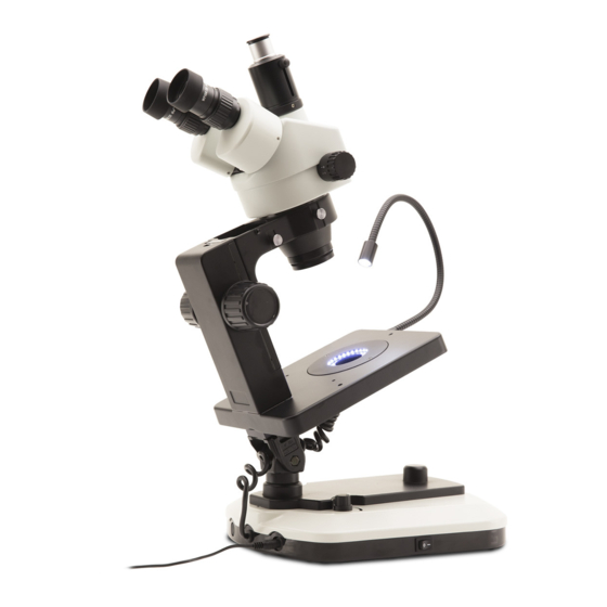
Instrument description OPTIGEM 10 EYEPIECE DIOPTER COMPENSATION RING MICROSCOPE BODY KOOM KNOB FOCUS KNOB INCIDENT LIGHT ILLUMINATOR WITH FLEXIBLE ARM ON/OFF - LIGHT INTENSITY ADJUSTMENT KNOB SUPPORT FOR ILLUMINATION HORIZONTAL SELECTOR KNOB OBSERVATION MAIN SWITCH Page 7... - Page 8 Opposite side DARK FIELD ILLUMINATOR JEWEL CLIP VELVET DARKFIELD SLIDER TRANSMITTED LIGHT ILLUMINATOR TILTING KNOB Page 8...
-
Page 9: Optigem
OPTIGEM 20 EYEPIECE DIOPTER COMPENSATION RING PHOTO/TV PORT MICROSCOPE BODY KOOM KNOB FOCUS KNOB INCIDENT ILLUMINATOR WITH FLEXIBLE ARM ON/OFF - LIGHT INTENSITY ADJUSTMENT KNOB SUPPORT FOR ILLUMINATION HORIZONTAL SELECTOR KNOB OBSERVATION MAIN SWITCH Page 9... - Page 10 Opposite side DARK FIELD ILLUMINATOR JEWEL CLIP VELVET DARKFIELD SLIDER TRANSMITTED LIGHT ILLUMINATOR TILTING KNOB Page 10...
-
Page 11: Assembling
Assembling 1. Remove the base from its packaging and place it on a flat surface. The base is already assembled from the factory and does not require any further assembly procedure other than that of mounting the microscope body. 2. Insert the microscope body in the holder. (Fig. 1) F ig. - Page 12 6. Insert the jewel clip in one of the three threaded holes on the stage. (Fig. 5) ② F ig. 5 ig. 5 Installing the photo port (OPTIGEM-20) ① 1. Loosen the fixing screws ① of the supplied photo port and remove the existing photo port.
-
Page 13: Use Of The Microscope
Use of the microscope Adjusting interpupillary distance Hold the right and left eyepiece tube with both hands and adjust the interpupillary distance by moving the two parts until one circle of light can be seen. (Fig. 8) • If two circles appear, the interpupillary distance is too big. •... -
Page 14: Magnification
4. Adjust the diopter compensation ring of the right eyepiece ⑤ (Fig. 12) until the image of the right eyepiece is clear and ⑤ sharp. Repeat the procedure for the left eyepiece. 5. Check the focus of the image for the whole zoom range. It should be perfectly parfocal (focus is maintained during the change of magnification). -
Page 15: Use Of Illumination
Use of illumination The OPTIGEM microscope comes with an integrated LED illumi- nation system that consists of three separate light sources: 1. Incident illumination: through a flexible arm attached to the microscope stage. 2. Transmitted illumination: from the hole of the microscope stage. -
Page 16: Use Of The Stand In Horizontal Position
Use of the stand in horizontal position ② The OPTIGEM microscope stand can easily be tilted in order to observe objects in a horizontal position. 1. Loosen the head fixing knob (Fig. 2) and rotate the head by 180°. 2. Fix the screw again. 3. -
Page 17: Use Of Optional Accessories
10. Use of optional accessories A wide range of accessories are available for the OPTIGEM microscope: gemology clips, an iris aperture diaphragm, a polarization analysis kit, an optical unit to switch the dark field from a “sharp” (gem exhibition) to a “soft” (diamond analysis) mode and a translating cell holder with vacuum pick-up and quartz immersion cells complete and enhance the instrument usability. -
Page 18: Polarized Light Analysis
5. Switch on the LED ring illuminator (position “b”, Fig. 17). • If you want it is possible to put your gem directly on the dia- phragm, after closing it to a proper diameter. (Fig. 25) F ig. 25 ig. 25 10.2 Polarized light analysis •... -
Page 19: Immersion Analysis With Translating Holder
10.4 Immersion analysis with translating holder • ST-203 + ST-204 + ST-207 are needed. 1. Install ST-207 as described in chapter 10.1. (Fig. 29) F ig. 29 ig. 29 2. Stack translating holder ST-204 and fix it with another spac- er. -
Page 20: Immersion Analysis With Translating Holder And Vacuum Pick Up
5. Install the jewel clip on the vertical rod, in order to hold the sample inside the immersion cell. (Fig. 33) F ig. 33 ig. 33 10.5 Immersion analysis with translating holder and vac- uum pick up • ST-203 + ST-204 + ST-207 + ST-205 are needed. 1. - Page 21 4. Now rotate the microscope into horizontal position and put the immersion cell ST-203 on the holder’s plate. (Fig. 37) F ig. 37 ig. 37 Page 21...
-
Page 22: Microphotography
11. Microphotography 11.1 Installing the C-mount adapter 1. Loosen the clamping screw ① on the trinocular port and re- move the dust cap ②. (Fig. 38) ② ② ① ① F ig. 38 ig. 38 2. Screw the C-mount adapter ③ to the camera ④ and insert the round dovetail of the C-mount into the empty hole of the trinocular port (Fig. -
Page 23: Maintenance
12. Maintenance Microscopy environment This microscope is recommended to be used in a clean, dry and shock free environment with a temperature of 5°-40°C and a maximum relative humidity of 75 % (non condensing). Use a dehumidifier if needed. To think about when and after using the microscope •... -
Page 24: Troubleshooting
13. Troubleshooting Review the information in the table below to troubleshoot operating problems. PROBLEM CAUSE SOLUTION I. Optical Section: The illumination is ON, but the field of The plug is not connected to the illu- Connect the cable view is dark. mination The brightness is too low Adjust to a proper setting... -
Page 25: Equipment Disposal
Equipment disposal Art.13 Dlsg 25 July 2005 N°151. “According to directives 2002/95/EC, 2002/96/EC and 2003/108/EC relating to the reduc- tion in the use of hazardous substances in electrical and electronic equipment and waste disposal.” The basket symbol on equipment or on its box indicates that the product at the end of its useful life should be collected sep- arately from other waste. - Page 26 OPTIKA S.r.l. ® Via Rigla, 30 - 24010 Ponteranica (BG) - ITALY Tel.: +39 035.571.392 info@optikamicroscopes.com - www.optikamicroscopes.com OPTIKA Spain spain@optikamicroscopes.com OPTIKA USA usa@optikamicroscopes.com OPTIKA China china@optikamicroscopes.com OPTIKA India india@optikamicroscopes.com OPTIKA Central America camerica@optikamicroscopes.com...
- Page 27 Serie OPTIGEM MANUALE DI ISTRUZIONI Modello OPTIGEM-10 OPTIGEM-20 Ver. 1.0 2021...
- Page 28 Sommario Avvertenza Informazioni sulla sicurezza Contenuto della confezione OPTIGEM 10 OPTIGEM 20 Disimballaggio Uso previsto Simboli Descrizione dello strumento OPTIGEM 10 OPTIGEM 20 Assemblaggio Uso del microscopio Regolazione della distanza interpupillare Messa a fuoco Regolazione della tensione delle manopole di messa a fuoco Compensazione diottrica Ingrandimento Accensione dell’illuminazione...
-
Page 29: Avvertenza
Avvertenza Questo microscopio è uno strumento scientifico di alta precisione, progettato per durare a lungo con una minima manutenzione; la realizzazione è secondo i migliori standard ottici e meccanici, per poter essere utilizzato quotidianamente. Vi ricordiamo che questo manuale contiene informazioni importanti per la sicurezza e per la manutenzione dello strumento, e deve quindi essere messo a disposizione di coloro che lo utilizzeranno. -
Page 30: Contenuto Della Confezione
Contenuto della confezione OPTIGEM 10 ① ② ③ ⑤ ④ ⑥ ① Stativo del microscopio ④ Pinza per pietre ② Corpo microscopio binoculare ⑤ Copertina antipolvere ③ Oculari ⑥ Alimentatore Pagina 30... -
Page 31: Optigem
OPTIGEM 20 ① ② ③ ⑤ ④ ⑥ ① Stativo del microscopio ④ Pinza per pietre ② Corpo microscopio trinoculare ⑤ Copertina antipolvere ③ Oculari ⑥ Alimentatore Pagina 31... -
Page 32: Disimballaggio
Disimballaggio Il microscopio è riposto in un imballo di polistirolo espanso. Rimuovere il nastro adesivo dal collo ed aprire la parte superiore dell’imballo. Fare attenzione a non far cadere le parti ottiche (obiettivi e oculari) nell’estrarre il microscopio dalla scatola per evitare che vengano danneggiati. -
Page 33: Descrizione Dello Strumento
Descrizione dello strumento OPTIGEM 10 OCULARE ANELLO REGOLAZIONE DIOTTRICA CORPO MICROSCOPIO MANOPOLA ZOOM MANOPOLA MESSA A ILLUMINATORE LUCE FUOCO INCIDENTE CON BRACCIO FLESSIBILE MANOPOLA ON/OFF - REGOLAZIONE INTENSITÀ LUMINOSA SUPPORTO PER MANOPOLA OSSERVAZIONE SELEZIONE ORIZZONTALE ILLUMINAZIONE INTERRUTTORE GENERALE Pagina 33... - Page 34 Lato opposto ILLUMINATORE CAMPO SCURO PINZA PER PIETRE SLITTA CAMPO SCURO VELLUTATO ILLUMINATORE LUCE TRASMESSA MANOPOLA PER INCLINAZIONE Pagina 34...
-
Page 35: Optigem
OPTIGEM 20 OCULARE ANELLO REGOLAZIONE DIOTTRICA USCITA FOTO/TV CORPO MICROSCOPIO MANOPOLA ZOOM MANOPOLA MESSA A ILLUMINATORE LUCE FUOCO INCIDENTE CON BRACCIO FLESSIBILE MANOPOLA ON/OFF - REGOLAZIONE INTENSITÀ LUMINOSA SUPPORTO PER MANOPOLA OSSERVAZIONE SELEZIONE ORIZZONTALE ILLUMINAZIONE INTERRUTTORE GENERALE Pagina 35... - Page 36 Lato opposto ILLUMINATORE CAMPO SCURO PINZA PER PIETRE SLITTA CAMPO SCURO VELLUTATO ILLUMINATORE LUCE TRASMESSA MANOPOLA PER INCLINAZIONE Pagina 36...
-
Page 37: Assemblaggio
Assemblaggio 1. Rimuovere la base dal suo imballo e posizionarla su una su- perficie piana. La base arriva già montata dalla fabbrica e non necessita di ulteriore procedura di montaggio, se non quella del montaggio del sistema di messa a fuoco. 2. - Page 38 6. Inserire la pinzetta per pietre in uno dei tre fori filettati sul tavolino. (Fig. 5) ② F ig. 5 ig. 5 Installare l’uscita foto (OPTIGEM-20) ① 1. Allentare le viti di fissaggio ① dell’uscita foto in dotazione e rimuovere l’uscita foto esistente. (Fig. 6) ①...
-
Page 39: Uso Del Microscopio
Uso del microscopio Regolazione della distanza interpupillare Afferrare con entrambe le mani i portaoculari destro e sinistro e regolare la distanza interpupillare spostando i tubi fino a che si osserva una sola immagine (Fig. 8). • Se si osservano due immagini la distanza è troppo elevata. •... -
Page 40: Ingrandimento
4. Regolare l’anello di regolazione diottrica dell’oculare destro ⑤ (Fig. 12) fino a che l’immagine è a fuoco. Ripetere la pro- ⑤ cedura per l’oculare sinistro. 5. Ora verificare la messa a fuoco del campione lungo l’intero range di zoom. Il sistema ora è perfettamente parafocale (il fuoco è... -
Page 41: Uso Dell'illuminazione
Uso dell’illuminazione Il microscopio OPTIGEM è dotato di un sistema di illuminazione a LED integrato che consiste in tre fonti di luce separate: 1. Illuminazione incidente: attraverso un braccio flessibile colle- gato al tavolino del microscopio. 2. Illuminazione trasmessa: dal foro del tavolino del microsco- pio. -
Page 42: Uso Dello Stativo In Posizione Orizzontale
Uso dello stativo in posizione orizzontale ② Lo stativo del microscopio OPTIGEM può essere facilmente in- clinato per osservare gli oggetti in posizione orizzontale. 1. Allentare la manopola di fissaggio della testa (Fig. 2) e ruo- tare la testa di 180°. 2. -
Page 43: Uso Degli Accessori Opzionali
10. Uso degli accessori opzionali Una vasta gamma di accessori è disponibile per il microscopio OPTIGEM: clip per gemmologia, un diaframma di apertura ad iride, un kit per l’analisi in polarizzazione, un’unità ottica per commutare il campo scuro da una modalità “forte” (analisi di gemme) a una “dolce”... -
Page 44: Analisi In Luce Polarizzata
5. Accendere l’illuminatore anulare a LED (posizione “b”, Fig. 17). • Se volete è possibile mettere la vostra gemma direttamente sul diaframma, dopo averlo chiuso ad un diametro adeguato. (Fig. 25) F ig. 25 ig. 25 10.2 Analisi in luce polarizzata •... -
Page 45: Analisi Ad Immersione Con Supporto Traslatore
10.4 Analisi ad immersione con supporto traslatore • Richiede ST-203 + ST-204 + ST-207. 1. Installare l’ST-207 come descritto al capitolo 10.1. (Fig. 29) F ig. 29 ig. 29 2. Impilare il supporto traslatore ST-204 e fissarlo con un altro distanziatore. -
Page 46: Analisi Ad Immersione Con Supporto Traslatore E Prelievo A Vuoto
5. Installare la pinza per pietre sull’asta verticale, per tenere il campione all’interno della cella d’immersione. (Fig. 33) F ig. 33 ig. 33 10.5 Analisi ad immersione con supporto traslatore e prelievo a vuoto • Richiede ST-203 + ST-204 + ST-207 + ST-205. 1. - Page 47 4. Ora ruotare il microscopio in posizione orizzontale e mettere la cella a immersione ST-203 sul piatto del supporto. (Fig. F ig. 37 ig. 37 Pagina 47...
-
Page 48: Microfotografia
11. Microfotografia 11.1 Uso di telecamere a passo “C” 1. Allentare la vite di bloccaggio ① sul tubo trinoculare e rimuovere il tappo antipolvere ②. (Fig. 38) ② ② ① ① F ig. 38 ig. 38 2. Avvitare l’adattatore passo C ③ alla telecamera ④ e installare l’attacco rotondo del passo C nel foro vuoto del tubo trinoculare, quindi riavvitare la vite di serraggio ①. -
Page 49: Manutenzione
12. Manutenzione Ambiente di lavoro Si consiglia di utilizzare il microscopio in un ambiente pulito e secco, privo di urti, ad una temperatura fra 0°C e 40°C e con una umidità relativa massima dell’85% (in assenza di condensazione). Si consiglia l’uso di un deumidificatore se necessario. Prima e dopo l’utilizzo del microscopio •... -
Page 50: Guida Alla Risoluzione Dei Problemi
13. Guida alla risoluzione dei problemi Consultare le informazioni riportate nella tabella seguente per risolvere eventuali problemi operativi. PROBLEMA CAUSA SOLUZIONE I. Sezione Ottica: Il LED è acceso, ma il campo visivo è L’alimentatore è scollegato. Collegarlo scuro La luminosità è troppo bassa Regolarla ad un livello adeguato I bordi del campo visivo sono vignettati L’illuminatore per luce incidente non è... -
Page 51: Smaltimento
Smaltimento Ai sensi dell’articolo 13 del decreto legislativo 25 luglio 2005 n°151. “Attuazione delle direttive 2002/95/CE, 2002/96/CE e 2003/108/CE, relative alla riduzione dell’uso di sostanze pericolose nelle apparecchiature elettriche ed elettroniche, nonché allo smaltimento dei rifiuti”. Il simbolo del cassonetto riportato sulla apparecchiatura o sulla sua confezione indica che il prodotto alla fine della propria vita utile deve essere raccolto separatamente degli altri rifiuti. - Page 52 OPTIKA S.r.l. ® Via Rigla, 30 - 24010 Ponteranica (BG) - ITALY Tel.: +39 035.571.392 info@optikamicroscopes.com - www.optikamicroscopes.com OPTIKA Spain spain@optikamicroscopes.com OPTIKA USA usa@optikamicroscopes.com OPTIKA China china@optikamicroscopes.com OPTIKA India india@optikamicroscopes.com OPTIKA Central America camerica@optikamicroscopes.com...
- Page 53 Serie OPTIGEM MANUAL DE INSTRUCCIONES Modelo OPTIGEM-10 OPTIGEM-20 Ver. 1.0 2021...
- Page 54 Índice Advertencia Información de seguridad Contenido del embalaje OPTIGEM 10 OPTIGEM 20 Desembalaje Utilización Símbolos Descripción del instrumenton OPTIGEM 10 OPTIGEM 20 Assembling Uso del microscopio Adjusting interpupillary distance Enfoque Ajuste de la tensión de los botones de enfoque Compensación dioptrica Aumento Encender la iluminación Uso de la iluminación...
-
Page 55: Advertencia
Advertencia Este microscopio es un instrumento científico de precisión. Su utilización está pensada para una larga duración con un mínimo nivel de mantenimiento. Para su fabricación se han utilizado elementos ópticos y mecánicos de elevada calidad que lo convierten en el instrumento ideal para la utilización diaria en las aulas y el laboratorio. Informamos que esta guía contiene importantes informaciones sobre la seguridad y el mantenimiento del producto y por lo tanto debe ser accesible a todos aquellos que utilizan dicho instrumento. -
Page 56: Contenido Del Embalaje
Contenido del embalaje OPTIGEM 10 ① ② ③ ⑤ ④ ⑥ ① Estativo del microscopio ④ Pinza para joyas ② Cuerpo microscopio binocular ⑤ Cubierta antipolvo ③ Oculares ⑥ Fuente de alimentación Página 56... -
Page 57: Optigem
OPTIGEM 20 ① ② ③ ⑤ ④ ⑥ ① Estativo del microscopio ④ Pinza para joyas ② Cuerpo microscopio trinocular ⑤ Cubierta antipolvo ③ Oculares ⑥ Fuente de alimentación Página 57... -
Page 58: Desembalaje
Desembalaje El microscopio esta embalado dentro de una caja de porexpan. Quitar el precinto que hay alrededor de la caja y abrirla. Tenga cuidado al abrir la caja ya que algunos accesorios ópticos como objetivos y oculares podrían caerse o dañarse. Con las dos manos (una sujetando el brazo y la otra la base) extraer el microscopio de dentro la caja de porexpan y poner sobre la mesa, procurando que ésta sea fuerte y estable. -
Page 59: Descripción Del Instrumenton
Descripción del instrumento OPTIGEM 10 OCULAR ANILLO DE COMPENSACIÓN DIÓPTRICA CUERPO MICROSCOPIO MANDO ZOOM MANDO DE ILUMINADOR DE ENFOQUE LUZ INCIDENTE CON BRAZO FLEXIBLE BOTÓN ENCENDIDO/ APAGADO - AJUSTE DE LA INTENSIDAD DE LA SOPORTE PARA BOTÓN SELECTOR OBSERVACIÓN DE ILUMINACIÓN HORIZONTAL INTERRUPTOR PRINCIPAL... - Page 60 Lado opuesto ILUMINADOR CAMPO OSCURO PINZA PARA JOYAS DESLIZADOR DE CAMPO OSCURO ILUMINADOR LUZ DE TERCIOPELO TRANSMITIDA POMO DE INCLINACIÓN Página 60...
-
Page 61: Optigem
OPTIGEM 20 OCULAR ANILLO DE COMPENSACIÓN DIÓPTRICA PUERTO DE FOTO/TV CUERPO MICROSCOPIO MANDO ZOOM MANDO DE ILUMINADOR DE ENFOQUE LUZ INCIDENTE CON BRAZO FLEXIBLE BOTÓN ENCENDIDO/ APAGADO - AJUSTE DE LA INTENSIDAD DE LA SOPORTE PARA BOTÓN SELECTOR OBSERVACIÓN DE ILUMINACIÓN HORIZONTAL INTERRUPTOR PRINCIPAL... - Page 62 Lado opuesto ILUMINADOR CAMPO OSCURO PINZA PARA JOYAS DESLIZADOR DE CAMPO OSCURO DE TERCIOPELO ILUMINADOR LUZ TRANSMITIDA POMO DE INCLINACIÓN Página 62...
- Page 63 Montaje 1. Retire la base de su embalaje y colóquela sobre una superfi- cie plana. La base ya viene montada de fábrica y no requiere ningún otro procedimiento de montaje que no sea el de mon- tar el cuerpo del microscopio. 2.
- Page 64 6. Inserte el clip para joyas en uno de los tres orificios roscados de la platina. (Fig. 5) ② F ig. 5 ig. 5 Instalación de la salida foto (OPTIGEM-20) ① 1. Afloje los tornillos de fijación ① de la salida de foto suminis- trada y retire la salida de foto existente.
-
Page 65: Uso Del Microscopio
Uso del microscopio Ajustar la distancia interpupilar Sostener el tubo del ocular derecho e izquierdo con ambas ma- nos y ajustar la distancia interpupilar moviendo las dos partes hasta que se pueda ver un círculo de luz. (Fig. 8) • Si miras dos imágenes, la distancia es demasiado grande. -
Page 66: Aumento
4. Ajustar el anillo de compensación de dioptrías del ocular de- recho ⑤ (Fig. 12) hasta que la imagen del ocular derecho ⑤ sea clara y nítida. Repetir el procedimiento para el ocular izquierdo. 5. A continuación, comprobar el enfoque de la imagen para todo el rango de zoom. -
Page 67: Uso De La Iluminación
Uso de la iluminación El microscopio OPTIGEM viene con un sistema de iluminación LED integrado que consta de tres fuentes de luz independientes: 1. Iluminación incidente: a través de un brazo flexible unido a la platina del microscopio. 2. Iluminación transmitida: desde el orificio de la platina del mi- croscopio. -
Page 68: Uso Del Soporte En Posición Horizontal
Uso del soporte en posición horizontal ② El soporte del microscopio OPTIGEM puede inclinarse fácilmen- te para observar objetos en posición horizontal. 1. Aflojar el pomo de fijación del cabezal (Fig. 2) y girar el ca- bezal 180°. 2. Fijar el tornillo de nuevo. 3. -
Page 69: Uso De Accesorios Opcionales
10. Uso de accesorios opcionales Existe una amplia gama de accesorios para el microscopio OPTIGEM: pinzas gemológicas, un diafragma de apertura del iris, un kit de análisis de polarización, una unidad óptica para cambiar el campo oscuro de un modo “nítido” (exposición de gemas) a uno “suave”... -
Page 70: Análisis De Luz Polarizada
5. Encender el anillo luminoso LED (posición “b”, Fig. 17). • Si lo desea, es posible colocar su gema directamente sobre el diafragma, después de cerrarlo a un diámetro adecuado. (Fig. 25) F ig. 25 ig. 25 10.2 Análisis de luz polarizada •... -
Page 71: Análisis En Inmersión Con Soporte De Traslación
10.4 Análisis en inmersión con soporte de traslación • Se necesitan ST-203 + ST-204 + ST-207. 1. Instalar el ST-207 como se describe en el capítulo 10.1. (Fig. F ig. 29 ig. 29 2. Apilar el soporte de traslación ST-204 y fijarlo con otro espa- ciador. -
Page 72: Análisis En Inmersión Con Soporte De Traslación Y Toma De Vacío
5. Instalar el clip de joya en la varilla vertical, con el fin de man- tener la muestra dentro de la célula de inmersión. (Fig. 33) F ig. 33 ig. 33 10.5 Análisis en inmersión con soporte de traslación y toma de vacío •... - Page 73 4. Ahora girar el microscopio en posición horizontal y poner la célula de inmersión ST-203 en la placa del soporte. (Fig. 37) F ig. 37 ig. 37 Página 73...
-
Page 74: Microfotografía
11. Microfotografía 11.1 Uso de cámaras de paso “C” 1. Aflojar el tornillo ① del tubo trinocular y quitar la tapa negra ②. (Fig. 38) ② ② ① ① F ig. 38 ig. 38 2. Colocar el adaptador paso C a la cámara ④ e insertar el conjunto sobre el puerto trinocular, luego sujetarlo con el tornillo para que no se caiga ①. -
Page 75: Mantenimiento
12. Mantenimiento Ambiente de trabajo Se aconseja utilizar este microscopio en un ambiente limpio y seco; también se deben evitar los impactos. La temperatura de trabajo recomendada es de 0-40°C y la humedad relativa máxima es de 85 % (en ausencia de condensación). Si es necesario, utilizar un deshumidificador. -
Page 76: Guía De Solución De Problemas
13. Guía de solución de problemas Revisar la información en la tabla a continuación para solucionar problemas de funcionamiento. PROBLEMA CAUSA SOLUCIÓN I. Sección Óptica: El iluminador está encendido, pero el El enchufe no está conectado al siste- Conectar campo visible está oscuro ma de iluminación La luminosidad es demasiado baja Regular la luminosidad... -
Page 77: Medidas Ecológicas Y Reciclaje
Medidas ecológicas y reciclaje De conformidad con el artículo 13 del Decreto Legislativo Nº 151, de 25 de julio de 2005. “Aplicación de las Directivas 2002/95/CE, 2002/96/CE y 2003/108/CE sobre la reducción del uso de sustancias peligrosas en aparatos eléctricos y elec- trónicos y la eliminación de residuos. - Page 78 OPTIKA S.r.l. ® Via Rigla, 30 - 24010 Ponteranica (BG) - ITALY Tel.: +39 035.571.392 info@optikamicroscopes.com - www.optikamicroscopes.com OPTIKA Spain spain@optikamicroscopes.com OPTIKA USA usa@optikamicroscopes.com OPTIKA China china@optikamicroscopes.com OPTIKA India india@optikamicroscopes.com OPTIKA Central America camerica@optikamicroscopes.com...
- Page 79 Série OPTIGEM MANUEL D’UTILISATION Modèle OPTIGEM-10 OPTIGEM-20 Ver. 1.0 2021...
- Page 80 Sommaire Avertissement Précautions Contenu de l’emballage OPTIGEM 10 OPTIGEM 20 Déballage Emploi prévu Symboles Description de l’instrument OPTIGEM 10 OPTIGEM 20 Assemblage Utilisation du microscope Ajuster la distance interpupillaire Mise au point Réglage de la tension des boutons de mise au point Compensation dioptrique Grossissement Allumer l’éclairage...
-
Page 81: Avertissement
Avertissement Le présent microscope est un appareil scientifique de précision créé pour offrir une durée de vie de plusieurs années avec un niveau d’entretien minimum. Les meilleurs composants optiques et mécaniques ont été utilisés pour sa conception ce qui fond de lui un appareil idéal pour une utilisation journalière. Ce guide contient des informations importantes sur la sécurité... -
Page 82: Contenu De L'emballage
Contenu de l’emballage OPTIGEM 10 ① ② ③ ⑤ ④ ⑥ ① Statif du microscope ④ Clip à bijoux ② Corps du microscope binoculaire ⑤ Cache-poussière ③ Oculaires ⑥ Alimentation électrique Page 82... - Page 83 OPTIGEM 20 ① ② ③ ⑤ ④ ⑥ ① Statif du microscope ④ Clip à bijoux ② Corps du microscope trinoculaire ⑤ Cache-poussière ③ Oculaires ⑥ Alimentation électrique Page 83...
-
Page 84: Déballage
Déballage Le microscope est emballé dans du polystyrène expansé. Enlever le ruban adhésif et retirer la partie supérieure de l’emballage. Retirer soigneusement le microscope et ses composants de l’emballage, utiliser les deux mains pour éviter de faire tomber et de casser les accessoires qu’il contient. L’appareil doit toujours être posé sur une surface stable, lisse et horizontale. -
Page 85: Description De L'instrument
Description de l’instrument OPTIGEM 10 OCULAIRE ANNEAU DE COMPENSATION DIOPTRIQUE CORPS DU MICROSCOPE BOUTON DE ZOOM BOUTON DE ILLUMINATEUR À MISE AU POINT LUMIÈRE INCIDENTE AVEC BRAS FLEXIBLE BOUTON ON/OFF - RÉGLAGE DE L’INTENSITÉ DE L’ÉCLAIRAGE SUPPORT POUR BOUTON DE L’OBSERVATION SÉLECTION DE HORIZONTALE... - Page 86 Côté opposé ILLUMINATEUR DE FOND NOIR CLIP À BIJOUX CURSEUR FOND NOIR EN VELOURS ILLUMINATEUR À LUMIÈRE TRANSMISE BOUTON INCLINABLE Page 86...
-
Page 87: Optigem
OPTIGEM 20 OCULAIRE ANNEAU DE COMPENSATION DIOPTRIQUE SORTIE PHOTO/TV CORPS DU MICROSCOPE BOUTON DE ZOOM BOUTON DE ILLUMINATEUR À MISE AU POINT LUMIÈRE INCIDENTE AVEC BRAS FLEXIBLE BOUTON ON/OFF - RÉGLAGE DE L’INTENSITÉ DE L’ÉCLAIRAGE SUPPORT POUR BOUTON DE L’OBSERVATION SÉLECTION DE HORIZONTALE L’ÉCLAIRAGE... - Page 88 Côté opposé ILLUMINATEUR DE FOND NOIR CLIP À BIJOUX CURSEUR FOND NOIR EN VELOURS ILLUMINATEUR À LUMIÈRE TRANSMISE BOUTON INCLINABLE Page 88...
-
Page 89: Assemblage
Assemblage 1. Retirer la base de son emballage et la placer sur une surface plane. La base est déjà assemblée en usine et ne nécessite aucune autre procédure d’assemblage que celle du montage du corps du microscope. 2. Insérer le corps du microscope dans le support. (Fig. 1) F ig. - Page 90 6. Insérer le clip à bijoux dans l’un des trois trous filetés de la platine. (Fig. 5) ② F ig. 5 ig. 5 Installer le port photo (OPTIGEM-20) ① 1. Desserrer les vis de fixation ① du port photo fourni et retirer le port photo existant.
-
Page 91: Utilisation Du Microscope
Utilisation du microscope Ajuster la distance interpupillaire Tenez le tube oculaire droit et gauche avec les deux mains et ajustez la distance interpupillaire en déplaçant les deux parties jusqu’à ce qu’un cercle de lumière soit visible. (Fig. 8) • Si deux cercles apparaissent, la distance interpupillaire est trop grande. -
Page 92: Grossissement
4. Régler la bague de compensation dioptrique de l’oculaire droit ⑤ (Fig. 12) jusqu’à ce que l’image de l’oculaire droit soit ⑤ nette. Répéter la procédure pour l’oculaire gauche. 5. Ensuite, vérifiez la mise au point de l’image pour toute la plage de zoom. -
Page 93: Utilisation De L'illumination
Utilisation de l’illumination Le microscope OPTIGEM est équipé d’un système d’éclairage LED intégré qui se compose de trois sources lumineuses dis- tincte: 1. Éclairage incident: par un bras flexible fixé à la platine du microscope. 2. Éclairage transmis: par le trou de la platine du microscope. 3. -
Page 94: Utilisation Du Support En Position Horizontale
Utilisation du support en position horizontale ② Le statif de microscope OPTIGEM peut facilement être incliné afin d’observer des objets en position horizontale. 1. Desserrez le bouton de fixation de la tête (Fig. 2) et faites pivoter la tête de 180°. 2. -
Page 95: Utilisation D'accessoires En Option
10. Utilisation d’accessoires en option Une large gamme d’accessoires est disponible pour le microscope OPTIGEM: des pinces de gemmologie, un diaphragme d’ouverture de l’iris, un kit d’analyse de polarisation, une unité optique pour commuter le fond noir d’un mode “net” (analyse de pierres précieuses) à... -
Page 96: Analyse En Lumière Polarisée
5. Allumer l’illuminateur annulaire à LED (position “b”, Fig. 17). • Si vous le souhaitez, il est possible de poser votre pierre pré- cieuse directement sur le diaphragme, après l’avoir fermé à un diamètre approprié. (Fig. 25) F ig. 25 ig. -
Page 97: Analyse Par Immersion Avec Support Translateur
10.4 Analyse par immersion avec support translateur • ST-203 + ST-204 + ST-207 sont nécessaires. 1. Installer le ST-207 comme décrit au chapitre 10.1. (Fig. 29) F ig. 29 ig. 29 2. Empiler le support de translation ST-204 et le fixer avec une autre entretoise. -
Page 98: Analyse Par Immersion Avec Support Translateur Et Prise De Vide
5. Installer la pince à bijoux sur la tige verticale, afin de main- tenir l’échantillon à l’intérieur de la cellule d’immersion. (Fig. F ig. 33 ig. 33 10.5 Analyse par immersion avec support translateur et prise de vide • ST-203 + ST-204 + ST-207 + ST-205 sont nécessaires. 1. - Page 99 4. Tourner maintenant le microscope en position horizontale et placer la cellule d’immersion ST-203 sur la plaque du sup- port.. (Fig. 37) F ig. 37 ig. 37 Page 99...
-
Page 100: Microphotographie
11. Microphotographie 11.1 Utilisation des caméras avec monture “C” 1. Desserrer la vis de fixation ① à la jointure du tube et enlever le couvercle de protection noir ②. (Fig. 38) ② ② ① ① F ig. 38 ig. 38 2. -
Page 101: Réparation Et Entretien
12. Réparation et entretien Environnement de travail Il est conseillé d’utiliser le microscope dans un environnement propre et sec, protégé des impactes, à une température comprise entre 0°C y 40°C et avec une humidité relative maximale de 85% (en absence de condensation). Il est conseillé d’utiliser un déshumidificateur si nécessaire. -
Page 102: Guide Résolution Des Problèmes
13. Guide résolution des problèmes Passer en revue les informations dans le tableau ci-dessous pour résoudre les problèmes opérationnels. PROBLÈME CAUSE SOLUTION I. Section Optique: La lampe est allumée mais le champ Les câbles d’alimentation ne sont Brancher les correctement visuel est sombre. -
Page 103: Ramassage
Ramassage Conformément à l’Article 13 du D.L du 25 Juillet 2005 nº151 Action des Directives 2002/95/CE, 2002/96/CE et 2003/108/CE, relatives à la réduction de l’utilisation de substances dan- gereuses dans l’appareil électrique et électronique et à l’élimination des résidus. Le Symbole du conteneur qui figure sur l’appareil électrique ou sur son emballage indique que le produit devra être, à la fin de sa vie utile, séparé... - Page 104 OPTIKA S.r.l. ® Via Rigla, 30 - 24010 Ponteranica (BG) - ITALY Tel.: +39 035.571.392 info@optikamicroscopes.com - www.optikamicroscopes.com OPTIKA Spain spain@optikamicroscopes.com OPTIKA USA usa@optikamicroscopes.com OPTIKA China china@optikamicroscopes.com OPTIKA India india@optikamicroscopes.com OPTIKA Central America camerica@optikamicroscopes.com...
- Page 105 Serie OPTIGEM BEDIENUNGSANLEITUNG Modell OPTIGEM-10 OPTIGEM-20 Ver. 1.0 2021...
- Page 106 Inhalt Hinweis Sicherheitsinformationen Verpackungsinhalt OPTIGEM 10 OPTIGEM 20 Auspacken Verwendung Wartung- und Gefahrzeichen Beschreibung des Instruments OPTIGEM 10 OPTIGEM 20 Montage Verwendung des mikroskops Einstellen des Augenabstandes Fokussierung Einstellen der Fokusspannung Dioptrienkompensation Vergrößerung Einschalten der Beleuchtung Verwendung der Beleuchtung Verwendung des Stativs in horizontaler Position Verwendung von optionalem Zubehör 10.1 Dunkelfeld-Analyse 10.2 Polarisierte Lichtanalyse...
-
Page 107: Hinweis
Hinweis Dieses Mikroskop ist ein wissenschaftliches Präzisionsgerät, es wurde entwickelt für eine jahrelange Verwendung bei einer minimalen Wartung. Dieses Gerät wurde nach den höchsten optischen und mechanischen Standards und zum täglichen Gebrauch hergestellt. Diese Bedienungsanleitung enthält wichtige Informationen zur korrekten und sicheren Benutzung des Geräts. -
Page 108: Verpackungsinhalt
Verpackungsinhalt OPTIGEM 10 ① ② ③ ⑤ ④ ⑥ ① Mikroskopstativ ④ Juwelenclip ② Binokularer Mikroskopkörper ⑤ Staubschutzhülle ③ Okulare ⑥ Netzteil Seite 108... - Page 109 OPTIGEM 20 ① ② ③ ⑤ ④ ⑥ ① Mikroskopstativ ④ Juwelenclip ② Trinokularer Mikroskopkörper ⑤ Staubschutzhülle ③ Okulare ⑥ Netzteil Seite 109...
-
Page 110: Auspacken
Auspacken Das Mikroskop ist in einer Schachtel aus Styroporschicht enthalten. Entfernen Sie das Klebeband von der Schachtel und öffnen Sie mit Vorsicht den oberen Teil, ohne Objektive und Okulare zu beschädigen. Mit beiden Händen (eine um dem Stativ und eine um der Basis) ziehen Sie das Mikroskop aus der Schachtel heraus und stellen Sie es auf eine stabile Ober- fläche. -
Page 111: Beschreibung Des Instruments
Beschreibung des Instruments OPTIGEM 10 OKULARE DIOPTRISCHER KOMPENSA- TIONSRING KÖRPERMIKROSKOP ZOOMSCHALTER FOKUSSIERKNOPF AUFLICHT-BELEUCHTUNG MIT FLEXIBLEM ARM EIN/AUS - EINSTELLKNOPF FÜR DIE LICHTSTÄRKE UNTERSTÜTZUNG FÜR HORIZONTALE BELEUCHTUNGSWAHLKNOPF BEOBACHTUNG HAUPTSCHALTER Seite 111... - Page 112 Gegenüberliegende Seite DUNKELFELD BELEUCHTUNG JUWELENCLIP SAMT-DUNKEL- FELD-SCHIEBER DURCHLICHTBE- LEUCHTUNG KIPPBARER KNOPF Seite 112...
- Page 113 OPTIGEM 20 OKULARE DIOPTRISCHER KOMPENSA- TIONSRING FOTO/TV AUSGANG KÖRPERMIKROSKOP ZOOMSCHALTER FOKUSSIERKNOPF AUFLICHT-BELEUCHTUNG MIT FLEXIBLEM ARM EIN/AUS - EINSTELLKNOPF FÜR DIE LICHTSTÄRKE UNTERSTÜTZUNG FÜR HORIZONTALE BELEUCHTUNGSWAHLKNOPF BEOBACHTUNG HAUPTSCHALTER Seite 113...
- Page 114 Gegenüberliegende Seite DUNKELFELD BELEUCHTUNG JUWELENCLIP SAMT-DUNKEL- FELD-SCHIEBER DURCHLICHTBE- LEUCHTUNG KIPPBARER KNOPF Seite 114...
-
Page 115: Montage
Montage 1. Nehmen die Basis aus der Verpackung und legen sie auf eine ebene Fläche. Die Basis ist bereits ab Werk zusammen- gebaut und erfordert außer der Montage des Mikroskopsta- tivs keine weiteren Montagearbeiten. 2. Einsetzen des Mikroskopkörpers in die Halterung. (Fig. 1) F ig. - Page 116 6. Stecken den Juwelenclip in eines der drei Gewindelöcher auf dem Tisch. (Fig. 5) ② F ig. 5 ig. 5 Installation des Fotoports (OPTIGEM-20) ① 1. Lösen die Befestigungsschrauben ① des mitgelieferten Fo- toports und entfernen den vorhandenen Fotoports. (Fig. 6) ①...
-
Page 117: Verwendung Des Mikroskops
Verwendung des mikroskops Einstellen des Augenabstandes Halten den rechten und linken Okulartubus mit beiden Händen und stellen den Augenabstand ein, indem die beiden Teile so lange bewegen, bis ein Lichtkreis zu sehen ist. (Fig. 8) • Wenn zwei Kreise erscheinen, ist der Augenabstand zu groß. •... -
Page 118: Vergrößerung
4. Stellen den Dioptrienausgleichsring des rechten Okulars ⑤ (Fig. 12) ein, bis das Bild des rechten Okulars klar und scharf ⑤ ist. Wiederholen den Vorgang für das linke Okular. 5. Überprüfen Sie nun den Fokus deie Probe über den gesam- ten Zoombereich. -
Page 119: Verwendung Der Beleuchtung
Verwendung der Beleuchtung Das OPTIGEM-Mikroskop ist mit einem integrierten LED-Be- leuchtungssystem ausgestattet, das aus drei separaten Licht- quellen besteht: 1. Auflichtbeleuchtung: durch einen flexiblen Arm, der am Mik- roskoptisch befestigt ist. 2. Durchlichtbeleuchtung: durch die Öffnung des Mikroskopti- sches. 3. Dunkelfeld-Beleuchtung: ein LED-Ring im Innern der Tisch- öffnung. -
Page 120: Verwendung Des Stativs In Horizontaler Position
Verwendung des Stativs in horizontaler Position ② Das OPTIGEM-Mikroskopstativ kann leicht gekippt werden, um Objekte in einer horizontalen Position zu beobachten. 1. Lösen den Kopfbefestigungsknopf (Fig. 2) und drehen den Kopf um 180°. 2. Befestigen die Schraube wieder. 3. Lösen den Kippknopf des Ständers ① (Fig. 20) und drehen den Ständer um 180°... -
Page 121: Verwendung Von Optionalem Zubehör
10. Verwendung von optional Zubehör Für das OPTIGEM-Mikroskop ist eine breite Palette an Zubehör erhältlich: Gemmologie-Clips, eine Irisblende, ein Polarisa- tionsanalyse-Kit, eine optische Einheit zum Umschalten des Dunkelfelds von einem “scharfen” (Edelsteinausstellung) auf einen “weichen” Modus (Diamantanalyse) und ein beweglicher Küvettenhalter mit Vakuumaufnahme und Quarz-Immersi- onsküvetten vervollständigen und verbessern die Nutzbarkeit des Instruments. -
Page 122: Polarisierte Lichtanalyse
5. Schalten die LED-Ringbeleuchtung ein (Position “b”, Fig. 17). • Wenn Sie möchten, können Sie Ihren Edelstein direkt auf die Membran legen, nachdem Sie sie auf einen angemessenen Durchmesser gebracht haben. (Fig. 25) F ig. 25 ig. 25 10.2 Polarisierte Lichtanalyse •... -
Page 123: Immersionsanalyse Mit Translatorischem Halter
10.4 Immersionsanalyse mit translatorischem Halter • ST-203 + ST-204 + ST-207 sind erforderlich. 1. Installieren ST-207 wie in Kapitel 10.1 beschrieben. (Fig. 29) F ig. 29 ig. 29 2. Übersetzungshalter ST-204 stapeln und mit einem weiteren Abstandshalter fixieren. (Fig. 30) F ig. -
Page 124: Immersionsanalyse Mit Translatorischer Halter Und Vakuumaufnahme
5. Befestigen die Juwelenclip an der vertikalen Stange, um die Probe in der Tauchzelle zu halten. (Fig. 33) F ig. 33 ig. 33 10.5 Immersionsanalyse mit translatorischer Halter und Vakuumaufnahme • ST-203 + ST-204 + ST-207 + ST-205 sind erforderlich. 1. - Page 125 4. Drehen nun das Mikroskop in die horizontale Position und setzen die Immersionszelle ST-203 auf die Platte des Hal- ters.. (Fig. 37) F ig. 37 ig. 37 Seite 125...
-
Page 126: Mikrofotografie
11. Mikrofotografie 11.1 Verwendung von C-Mount Kameras 1. Lösen Sie die Sicherungsschraube ① am Binokulartubus und entfernen Sie die Staubkappe ②. (Fig. 79) ② ② ① ① F ig. 38 ig. 38 2. Schrauben Sie den Adapterschritt “C” ③ an die Kamera ④ und montieren Sie die runde Halterung der Stufe C in die leere Bohrung des Binokulartubus (Fig. -
Page 127: Wartung
12. Wartung Arbeitsumfeld Es wird empfohlen, das Mikroskop an einem sauberen, trockenen und stoßsicheren Ort zu verwenden, bei einer Tempe- ratur zwischen 0° und 40° und einer Feuchtigkeit nicht über 85% (ohne Kondensation). Wenn nötig wird die Verwendung eines Luftentfeuchters empfohlen. Vor und nach dem Gebrauch des Mikroskops •... -
Page 128: Probleme Und Lösungen
13. Probleme und Lösungen Lesen Sie die Informationen in der folgenden Tabelle, um Probleme bei der Bedienung zu beheben. PROBLEM URSACHE LÖSUNG I. Optisches System: Die Beleuchtung ist eingeschaltet, Stromversorgungsstecker sind nicht Verbinden sie aber das Sichtfeld ist dunkel gut angeschlossen. Die Helligkeit ist zu gering. -
Page 129: Wiederverwertung
Wiederverwertung Gemäß dem Artikel 13 vom Dekret Nr. 151 vom 25.07.2005 “Umsetzung der Richtlinien 2002/95/EG, 2002/96/EG und 2003/108/EG in Bezug auf die Verwendung gefährlicher Stoffe in elektrischen und elektronischen Geräten sowie die Ab- fallentsorgung”. Das Symbol vom Müllcontainer erscheint auf dem Gerät oder der Verpackung und weist darauf hin, dass das Produkt Ende des Lebens separat von anderen Abfällen entsorgt werden muss. - Page 130 OPTIKA S.r.l. ® Via Rigla, 30 - 24010 Ponteranica (BG) - ITALY Tel.: +39 035.571.392 info@optikamicroscopes.com - www.optikamicroscopes.com OPTIKA Spain spain@optikamicroscopes.com OPTIKA USA usa@optikamicroscopes.com OPTIKA China china@optikamicroscopes.com OPTIKA India india@optikamicroscopes.com OPTIKA Central America camerica@optikamicroscopes.com...
- Page 131 Série OPTIGEM MANUAL DE INSTRUÇÕES Modelo OPTIGEM-10 OPTIGEM-20 Ver. 1.0 2021...
- Page 132 Tabela de Conteúdos Advertência Informações sobre a segurança Conteúdo da embalagem OPTIGEM 10 OPTIGEM 20 Desembalando Uso previsto Simbolos Descrição do instrument OPTIGEM 10 OPTIGEM 20 Montagem Uso do microscópio Adjusting interpupillary distance Focalização Ajuste da tensão de focagem Compensação dióptrica Ampliação Ligar a iluminação Uso da iluminação...
-
Page 133: Advertência
Advertência Este microscópio é um instrumento científico de alta precisão, projectado para durar um longo tempo com manutenção mínima; a sua realização respeita os melhores padrões ópticos e mecânicos, para que possa ser utilizado diariamente. Recordamos que este manual contém informações importantes para a segurança e a manutenção do instrumento, portanto deve ser colocado à... -
Page 134: Conteúdo Da Embalagem
Conteúdo da embalagem OPTIGEM 10 ① ② ③ ⑤ ④ ⑥ ① Suporte de microscópio ④ Clipe para jóias ② Corpo do microscópio binocular ⑤ Capa de pó ③ Oculares ⑥ Alimentação eléctrica Página 134... -
Page 135: Optigem
OPTIGEM 20 ① ② ③ ⑤ ④ ⑥ ① Suporte de microscópio ④ Clipe para jóias ② Corpo do microscópio trinocular ⑤ Capa de pó ③ Oculares ⑥ Alimentação eléctrica Página 135... -
Page 136: Desembalando
Desembalando O microscópio é alojado em um recipiente de isopor moldado. Remova a fita da borda do recipiente e levante a metade superior do recipiente. Tome algum cuidado para evitar que os itens ópticos (objetivas e oculares) cair e ficar danificado. Usando ambas as mãos (uma ao redor do braço e outra ao redor da base), levante o microscópio do recipiente e coloque-o em uma mesa estável. -
Page 137: Descrição Do Instrument
Descrição do instrumento OPTIGEM 10 OCULAR ANEL DE COMPENSAÇÃO DIÓPTRICA CORPO DO MICROSCÓPIO BOTÃO DE ZOOM BOTÃO DE ILUMINADOR DE LUZ FOCAGEM INCIDENTE COM BRAÇO FLEXÍVEL BOTÃO DE LIGAR/DESLIGAR - AJUSTE DA INTENSIDADE DA LUZ SUPORTE PARA BOTÃO SELECTOR OBSERVAÇÃO DE ILUMINAÇÃO HORIZONTAL INTERRUPTOR... - Page 138 Lado oposto ILUMINADOR DE CAMPO ESCURO CLIPE PARA JÓIAS DESLIZADOR DE CAMPO ESCURO ILUMINADOR DE DE VELUDO LUZ TRANSMITIDA BOTÃO DE INCLINAÇÃO Página 138...
-
Page 139: Optigem
OPTIGEM 20 OCULAR ANEL DE COMPENSAÇÃO DIÓPTRICA SAÍDA DE FOTO CORPO DO MICROSCÓPIO BOTÃO DE ZOOM BOTÃO DE ILUMINADOR DE LUZ FOCAGEM INCIDENTE COM BRAÇO FLEXÍVEL BOTÃO DE LIGAR/DESLIGAR - AJUSTE DA INTENSIDADE DA LUZ SUPORTE PARA BOTÃO SELECTOR OBSERVAÇÃO DE ILUMINAÇÃO HORIZONTAL INTERRUPTOR... - Page 140 Lado oposto ILUMINADOR DE CAMPO ESCURO CLIPE PARA JÓIAS DESLIZADOR DE CAMPO ESCURO DE VELUDO ILUMINADOR DE LUZ TRANSMITIDA BOTÃO DE INCLINAÇÃO Página 140...
-
Page 141: Montagem
Montagem 1. Retirar a base da sua embalagem e colocá-la sobre uma superfície plana. A base já está montada de fábrica e não re- quer qualquer outro procedimento de montagem para além da montagem do corpo do microscópio. 2. Inserir o corpo do microscópio no suporte. (Fig. 1) F ig. - Page 142 6. Inserir o clipe para jóias num dos três orifícios roscados da platina (Fig. 5) ② F ig. 5 ig. 5 Instalar a porta foto (OPTIGEM-20) ① 1. Desapertar os parafusos de fixação ① da saída de foto for- necida e remover a saída de foto existente. (Fig. 6) ①...
-
Page 143: Uso Do Microscópio
Uso do microscópio Ajuste da distância interpupilar Segurar os lados esquerdo e direito da cabeça de observação com ambas as mãos e ajustar a distância interpupilar rodando os dois lados até que um único círculo de luz seja visível. (Fig. 8) •... -
Page 144: Ampliação
4. Ajustar o anel de compensação de dioptrias da ocular direita ⑤ (Fig. 12) até que a imagem da ocular direita seja clara e ⑤ nítida. Repetir o procedimento para a ocular esquerda. 5. Verificar o foco da imagem para toda a gama de zoom. Deve ser perfeitamente parfocal (o foco é... -
Page 145: Uso Da Iluminação
Uso da iluminação O microscópio OPTIGEM vem com um sistema de iluminação LED integrado que consiste em três fontes de luz separadas: 1. Iluminação de incidências: através de um braço flexível liga- do à fase de microscópio. 2. Iluminação transmitida: a partir do orifício da platina do mi- croscópio. -
Page 146: Utilização Do Suporte Em Posição Horizontal
Utilização do suporte em posição horizontal ② O suporte de microscópio OPTIGEM pode ser facilmente inclina- do a fim de observar objectos em posição horizontal. 1. Soltar o botão de fixação da cabeça (Fig. 2) e rodar a cabeça em 180°. 2. -
Page 147: Uso De Acessórios Opcionais
10. Uso de acessórios opcionais Uma vasta gama de acessórios está disponível para o microscópio OPTIGEM: clipes de gemologia, um diafragma de abertura da íris, um kit de análise de polarização, uma unidade óptica para mudar o campo escuro de um modo “cortante” (exposição de gemas) para um modo “suave”... -
Page 148: Análise Em Luz Polarizada
5. Ligar o iluminador de anel de LED (posição “b”, Fig. 17). • Se quiser é possível colocar a sua gema directamente sobre o diafragma, após fechá-lo a um diâmetro adequado. (Fig. F ig. 25 ig. 25 10.2 Análise em luz polarizada •... -
Page 149: Análise Em Imersão Com Suporte De Translação
10.4 Análise em imersão com suporte de translação • ST-203 + ST-204 + ST-207 são necessários. 1. Instalar o ST-207 como descrito no capítulo 10.1. (Fig. 29) F ig. 29 ig. 29 2. Empilhar o suporte de translação ST-204 e fixá-lo com outro espaçador. -
Page 150: Análise Em Imersão Com Suporte De Translação E Captador De Vácuo
5. Instalar o clipe para jóias na haste vertical, a fim de segurar a amostra dentro da célula de imersão. (Fig. 33) F ig. 33 ig. 33 10.5 Análise em imersão com suporte de translação e captador de vácuo • ST-203 + ST-204 + ST-207 + ST-205 são necessários. - Page 151 4. Rodar agora o microscópio na posição horizontal e colocar a célula de imersão ST-203 na placa do suporte. (Fig. 37) F ig. 37 ig. 37 Página 151...
-
Page 152: Microfotografia
11. Microfotografia 11.1 Uso de câmaras de paso “C” 1. Desapertar o parafuso de aperto ① na porta trinocular e retirar a tampa do pó ②. (Fig. 38) ② ② ① ① F ig. 38 ig. 38 2. Aparafusar o adaptador C-mount ③ à câmara ④ e inserir o encaixe redondo do C-mount no orifício vazio da porta trinocular (Fig. -
Page 153: Manutenção
12. Manutenção Ambiente de trabalho Recomenda-se de utilizar o microscópio em um ambiente limpo e seco, sem o risco de colisões, a uma temperatura entre 0°C e 40°C e com uma humidade relativa máxima de 85% (em ausência de condensação). Recomenda-se o uso de um desumidificador, se necessário. -
Page 154: Resolução De Problemas
13. Resolução de problemas Reveja a informação na tabela abaixo para tentar solucionar problemas de operação. PROBLEMA CAUSA SOLUÇÃO I. Secção Óptica: A iluminação está ligada, mas o campo O plugue do suporte da lâmpada não Conecte-os de visão está escuro está... -
Page 155: Eliminação
Eliminação Art.13 Dlsg 25 de Julho de 2005 N°151. “De acordo com as Directivas 2002/95/CE, 2002/96/CE e 2003/108/CE relativas à redução do uso de substâncias perigosas em equipamentos eléctricos e electrónicos e à eliminação de resíduos. O símbolo do cesto no equipamento ou na sua caixa indica que o produto no final da sua vida útil deve ser recolhido sepa- radamente dos outros resíduos. - Page 156 OPTIKA S.r.l. ® Via Rigla, 30 - 24010 Ponteranica (BG) - ITALY Tel.: +39 035.571.392 info@optikamicroscopes.com - www.optikamicroscopes.com OPTIKA Spain spain@optikamicroscopes.com OPTIKA USA usa@optikamicroscopes.com OPTIKA China 100 Lauman Lane, Suite A, Hicksville, NY 11801 Tel: (877) 877-7274 | Fax: (516) 801-2046 china@optikamicroscopes.com Email: Info@nyscopes.com www.microscopeinternational.com...